
New research led by Dr Dorothea Böken and Prof Sir David Klenerman (UK DRI at Cambridge) reveals how the proteins associated with tau change as it forms increasingly large clumps in the brain. Published in Advanced Science, the study provides new insights into the molecular processes underlying Alzheimer's disease and highlights potential opportunities for targeting tau pathology at different stages of aggregation.
What was the challenge?
The build-up of abnormal tau protein in the brain is a key feature of Alzheimer's disease. Normally, tau helps maintain the internal structure of neurons, but in disease it can form small clumps, known as aggregates, which may eventually develop into larger fibres.
While these aggregates are closely linked to neurodegeneration, researchers still do not fully understand how they interact with other proteins in the brain. These interactions may influence how aggregates form, how cells respond to them and ultimately how they contribute to disease. Studying the smallest aggregates is particularly challenging, as they are difficult to detect using conventional techniques.
What did the team do and what did they find?
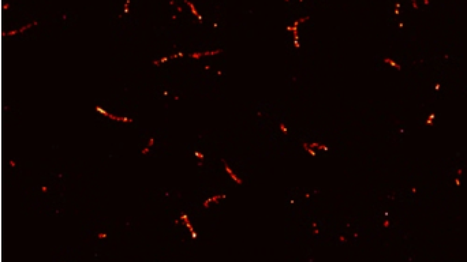
The researchers combined advanced protein analysis with highly sensitive techniques known as single-molecule methods to investigate how tau interacts with other proteins. While conventional methods typically measure large numbers of proteins together, single-molecule techniques allow researchers to detect and study individual protein molecules and tiny aggregates, which would otherwise be difficult to distinguish. This makes it possible to examine not only how much aggregated tau is present, but also the size of individual aggregates and which other proteins are associated with them.
Using donated brain tissue from people with Alzheimer's disease and healthy controls, the team identified proteins associated with different forms of tau, ranging from individual molecules to small aggregates and larger fibres. They then used single-molecule imaging to examine these interactions in more detail, alongside experiments in cultured nerve cells to investigate how they changed as tau aggregation progressed.
The team discovered that different forms of tau were associated with distinct groups of proteins involved in important cellular functions. Earlier forms of tau showed associations with proteins involved in cellular stress responses and protein quality control, while more mature aggregates were linked to proteins involved in energy metabolism, cell structure and communication between nerve cells. Crucially, by examining individual tau aggregates, the researchers could directly observe how the association of specific proteins differed depending on aggregate size.
Together, these findings suggest that tau aggregation involves a changing network of interactions with other cellular proteins, rather than simply the accumulation of tau itself.
We often think about Alzheimer's disease in terms of tau protein building up into harmful clumps. But our findings suggest that it is not just tau itself that matters, the other proteins associated with these clumps also change as they develop.
By understanding which proteins become involved at different stages, we hope to uncover new clues about how tau pathology affects nerve cells and identify opportunities to intervene in this process.
Postdoctoral Researcher
What is the impact?
Understanding how tau interacts with other proteins as aggregates develop could help researchers identify which cellular processes are affected at different stages of Alzheimer's pathology. These findings may ultimately point towards new therapeutic targets.
Importantly, the study also highlights the power of single-molecule techniques for understanding neurodegenerative diseases. By revealing tiny protein aggregates and their interactions that would otherwise be hidden within measurements of millions of molecules, these approaches offer a more detailed picture of the molecular changes occurring in the brain, potentially helping researchers identify early disease-related changes that conventional techniques might miss.
Reference: D. Böken, P. B. Lobo, Y. Wu, et al. “ Temporal Trajectories of the Tau Aggregate Interactome Reveal Stage-Specific Vulnerabilities in Alzheimer's Disease.” Advanced Science (2026): e77822. https://doi.org/10.1002/advs.77822
Banner image: Tiny clumps of tau protein, known as aggregates, visualised using super-resolution microscopy to reveal their different sizes and shapes. The tau aggregates were isolated from nerve cells grown in the laboratory. Credit: Dorothea Böken.