A new study led by Prof Adrian Isaacs (UK DRI at UCL) provides fresh insight into the early changes that occur in the brains of people with amyotrophic lateral sclerosis (ALS) and frontotemporal dementia (FTD), and reveals a response in cells that protects against neurodegeneration. The research, published in Nature Neuroscience, could identify new therapeutic avenues to treat the diseases.
What was the challenge?
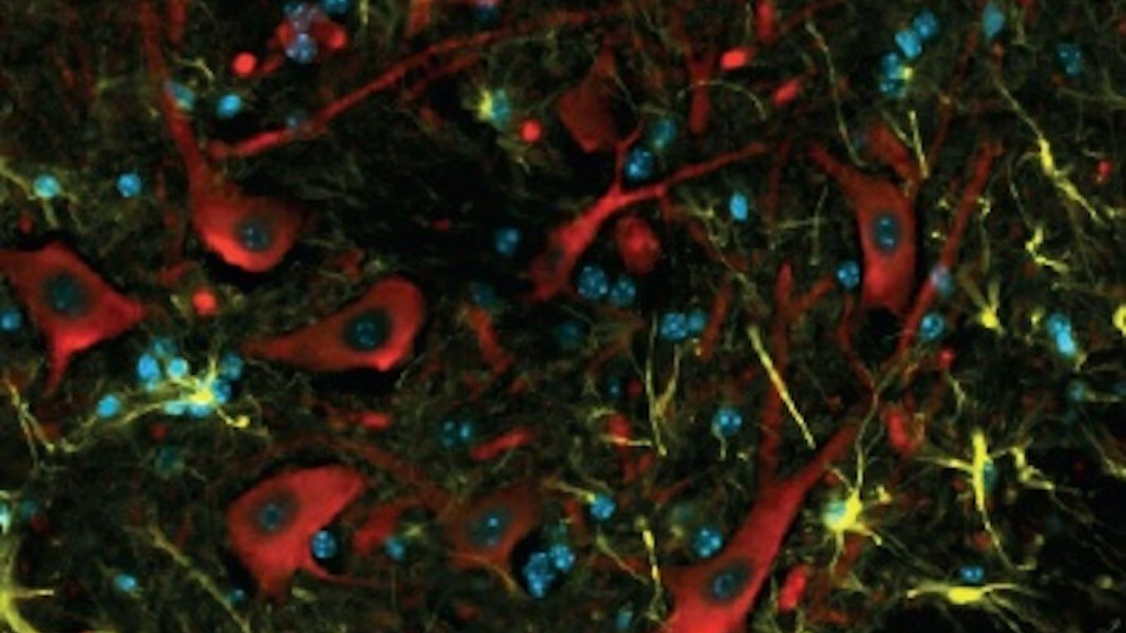
A mutation in a gene called C9orf72 is the most common cause of ALS and FTD. This faulty gene leads to the production of abnormal proteins which are a known feature of the diseases, but their impact has not yet been fully determined. In this study, the team set out to generate a new mouse model which more accurately recapitulated the pathology of the disease to examine the impact of these proteins in the brain and spinal cord.
We unexpectedly found that neurons can mount a neuroprotective effort early in the course of FTD and ALS. This provides important new information in our fight to understand and ultimately develop treatments for neurodegenerative diseases.Prof Adrian IsaacsUK DRI at UCL
What did the team do and what did they find?
With funding from the Motor Neurone Disease Association, the UK DRI and the Packard Center for ALS Research, the researchers created mouse models with a specific genetic makeup that mimicked features of ALS and FTD. When they studied the proteins found in the spinal cord of the mice, they discovered an increase in proteins related to the extracellular matrix , which usually provides structural support to cells – with levels of a protein called COL6A1 most raised.
Further investigation revealed that a protein called TGF-β1 played a key role in controlling this increase in extracellular matrix proteins. When the scientists experimented with this in human neurons grown in the lab, they found that the abnormal proteins generated by faulty C9orf72 triggered the production of TGF-β1 and COL6A1.
To understand the impact of these findings on the disease, the team conducted experiments in fruit flies and human neurons. They discovered that reducing TGF-β1 or COL6A1 caused neurodegeneration to worsen in fruit flies, while increasing these proteins protected human motor neurons from certain types of damage – suggesting that the proteins could be triggering a protective response in cells.
What was the impact of these findings?
The new model provides insight into early changes that occur in the brains of people with ALS and FTD. It could help to identify new therapeutic avenues to treat these diseases.
Prof Isaacs said:
“I am delighted that this collaborative effort has come to fruition. We unexpectedly found that neurons can mount a neuroprotective effort early in the course of FTD and ALS. This provides important new information in our fight to understand and ultimately develop treatments for neurodegenerative diseases.”
Co-senior author Prof Elizabeth Fisher, Professor of Neurogenetics at UCL, added:
“These new mouse models have taken many years to make and characterise, with funding from UK and USA, and they will greatly help our efforts to understand why motor neurons die as a result of mutation in the C9orf72 gene.”
To find out more about Prof Adrian Isaacs' work, visit his UK DRI profile. To keep up to date with the latest UK DRI news and events, sign up to receive our monthly newsletter.
Article published: 29 February 2024
Banner image credit: Mireia Carcolé