A new study by Prof Paul Matthews (UK DRI at Imperial) and colleagues reveals that a widely used marker of inflammation does not accurately reflect neurodegeneration in humans, as previously thought. The study is published in Nature Communications.
What was the challenge?
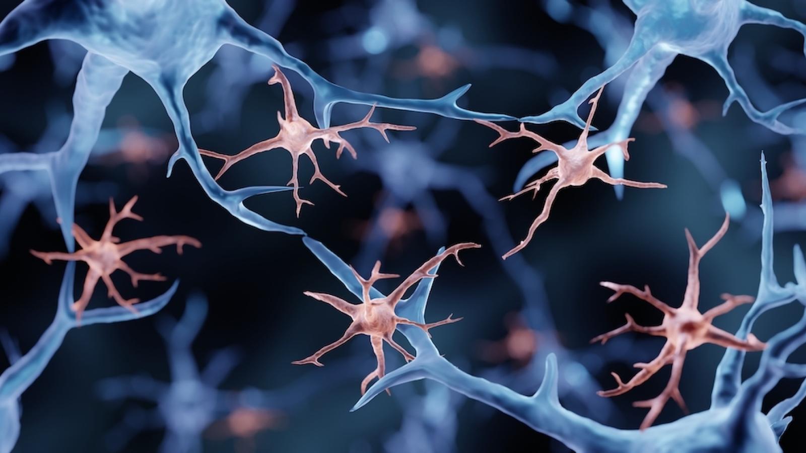
Positron emission tomography (PET) brain imaging targeting a protein known as translocator protein (TSPO) is widely used to measure neuroinflammation in humans, as it is believed to be an indicator that microglia, the brains resident immune cells, are activated. The team set out to test this hypothesis using human tissue and mouse models.
This paper provides a powerful example of how the UK DRI can pull together a large international group to provide definitive data that fundamentally changes old ways of thinking.Prof Paul MatthewsCentre Director of the UK DRI at Imperial
What did the team do?
The researchers analysed human brain tissue from donors with Alzheimer’s, amyotrophic lateral sclerosis and multiple sclerosis, and compared the TSPO expression with that of mouse models.
They found that, unlike in mice, human microglia do not increase TSPO expression in response to inflammation. In the human brain tissue analysed in the study, the researchers instead found more microglia than in control brain. Therefore, the team propose that the increased TSPO may indicate increased density of microglia, not activation as it does in mice.
What is the impact of these findings?
These findings fundamentally alter the interpretation of decades of studies that utilise TSPO PET imaging. The new study suggests that TSPO PET signal in humans provides an indication of an increased accumulation of microglia and other inflammatory cells (e.g. astrocytes), not activation of microglia.
Prof Matthews said:
“This paper provides a powerful example of how the UK DRI can pull together a large international group to provide definitive data that fundamentally changes old ways of thinking - in this case about imaging inflammation. I am excited particularly by the genomic discovery approach – first of its kind - leading to the identification of new, specific markers of microglial pro-inflammatory activation. Taking this to the next stages for their development will be transformative!”
Read more about this study in the full paper below or the preprint summary from Alzforum. Find out more about Prof Paul Matthews’ research by visiting his UK DRI profile.
Reference: Nutma, E., Fancy, N., Weinert, M. et al. Translocator protein is a marker of activated microglia in rodent models but not human neurodegenerative diseases. Nat Commun 14, 5247 (2023). https://doi.org/10.1038/s41467...
Article published: 8 September 2023
Banner image: Shutterstock/Artur Plawgo
