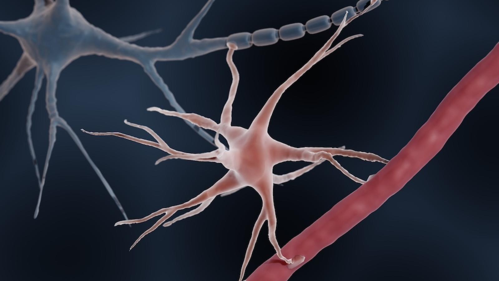
Researchers led by Dr Lorena Arancibia Cárcamo and Dr Anna Mallach at the UK DRI at UCL, the Francis Crick Institute and the VIB-KU Leuven Center for Brain & Disease Research have revealed how communication between support cells in the brain disrupts signals between neurons in mice modelling Alzheimer’s disease.
Understanding the messages between cells that accumulate next to amyloid plaques – one of the hallmarks of Alzheimer’s – could help researchers understand how the condition develops and how to treat it.
In research published this week in Cell Reports, scientists investigated the role of two support cells: astrocytes, which help neurons carry out their functions, and microglia, the resident immune cells in the brain.
Previous research has shown that both of these cells are involved in the development of Alzheimer’s, but how they interact with each other and with neurons was not fully understood.
The team used a state-of-the-art technique called spatial transcriptomics, which allows genetic signals to be mapped to different cell types and their location in the brain, showing how cells interact with each other.
We need to work out how to specifically target the signals astrocytes and microglia produce when found near amyloid plaques.Dr Lorena Arancibia CárcamoUK DRI at UCL & the Crick
They found that microglia built up near plaques all across the mouse brain, but astrocytes only accumulated next to plaques in certain regions, such as the hippocampus, an area associated with learning and memory.
Focusing on areas of amyloid plaques where both of these cells were accumulating, the team identified signals showing that microglia and astrocytes were talking to each other. The more microglia there were around the plaque, the more toxic to neurons the astrocytes became, leading to reduced brain activity.
They found this was because the ‘activated’ astrocytes disrupted nerve cell communication, by increasing a chemical messenger called GABA and decreasing another called glutamate.
As amyloid plaques (white) increase in severity, more microglia surround them, which in turn causes astrocytes (green) to become more toxic. Credit: Anna Mallach, the Francis Crick Institute.
Dr Anna Mallach, Postdoctoral Research Fellow in the Cellular Phase of Alzheimer’s Disease Laboratory at the Crick, and co-first author with Magdalena Zielonka, said:
“A very recent surge in these novel techniques has let us zoom in and see cell interactions around plaques in a resolution that hasn’t previously been possible.
“We knew that support cells, or glia, play an important role in Alzheimer’s disease, but this work has confirmed that microglia kick off the disease cascade, and directly communicate to astrocytes, which in turn disrupt signals between neurons.”
An untapped treatment possibility
The next step for the researchers is to investigate the proteins involved in this cell cross-talk and see if they can be blocked, shutting down this specific message between astrocytes and microglia.
Dr Lorena Arancibia Cárcamo, Principal Staff Scientist and Co-Leader of Prof Bart De Strooper's lab, UK DRI at UCL and the Crick, said:
“New amyloid-clearing drugs in the pipeline for Alzheimer’s disease are incredibly exciting but the role of microglia and astrocytes is also significant.
“Both of these cells would be useful targets for Alzheimer’s treatments. We need to work out how to specifically target the signals they produce when found near amyloid plaques in the cases of disease and understand if we can reduce the harmful effects on neurons.”
Source: The Francis Crick Institute
Article published: 5 June 2024
Banner image: Shutterstock/ART-ur
