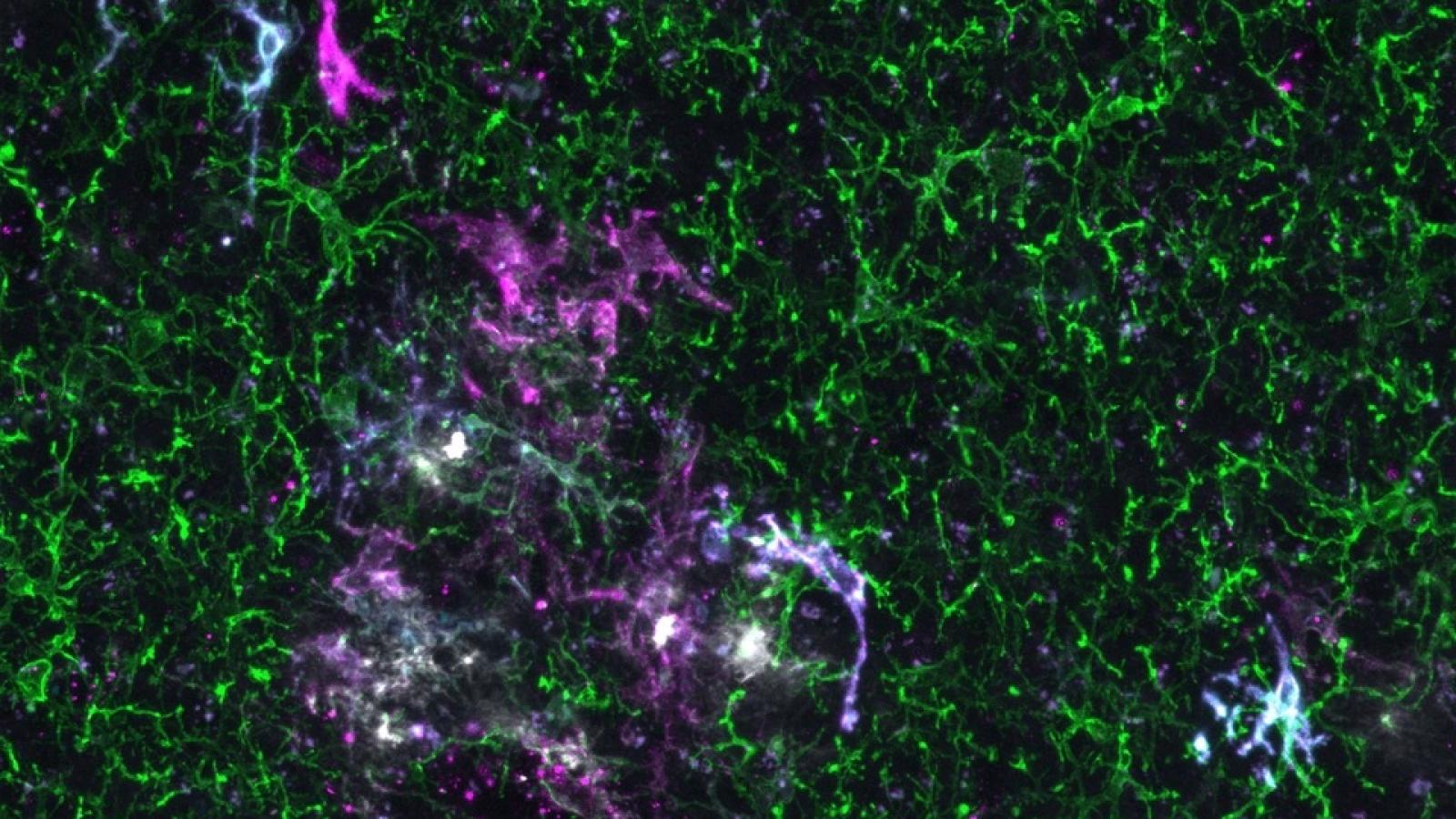
Microglia, the brain’s resident immune cells, are implicated in Alzheimer’s and other neurodegenerative diseases, but the exact mechanisms behind this are not yet understood due to the complexities involved in studying them in human brain samples. A team led by Prof Bart De Strooper (UK DRI at UCL and VIB-KU Leuven) and Prof Renzo Mancuso (VIB-UAntwerp) has transplanted human microglia into mouse brains to better observe how the microglia respond to the disease environment. Their findings, published in Nature Neuroscience, will help scientists understand the complex mechanisms involved in Alzheimer's disease.
Microglia are responsible for clearing debris and responding to inflammation in the brain. Scientists have been studying these cells in Alzheimer’s, as they play a central role in the disease, especially in the build-up of and early response to amyloid beta plaques, one of the major hallmarks of the disease. The microglia react to the plaques as they are perceived as foreign, driving neuroinflammation in the brain.
Our findings validate this xenograft model as a powerful tool to investigate the genetics underlying microglial response in Alzheimer's.Prof Bart De Strooper
Studying these cells in human brain samples post-mortem can be challenging because of genetic differences between people, the time between death and examination, and the presence of other brain disorder. These issues may contribute to the varying microglial reactions observed in postmortem brain samples. A further complication arises as it is not possible to test the effects of interventions like medication on post-mortem brains.
The first authors of the study, Dr Nicola Fattorelli and Dr Anna Martinez Muriana, together with their colleagues at the VIB-KU Leuven Center for Brain & Disease Research, the VIB-UAntwerp Center for Molecular Neurology and the UK DRI, developed a unique mouse model. This xenotransplantation model is genetically engineered to mimic the amyloid beta plaque accumulations seen in humans with Alzheimer’s and can be transplanted with stem-cell-derived human microglia. Previously, a similar model was able to show how transplanted human neurons die in Alzheimer’s. Now, this approach has allowed the researchers to investigate how human microglia respond to amyloid plaques during the course of the disease.
Microglial responses to Alzheimer’s
The scientists found that human microglia showed a much more complex immune response to amyloid beta than their rodent counterparts. Human microglia also displayed a different genetic transition from the normal to the reactive state of the cells.
“This could have implications for the development of treatments,” says Prof Renzo Mancuso, first author of the study and Group Leader at the VIB-UAntwerp Center for Molecular Neurology. “Researchers need to be cautious when using mouse models to study Alzheimer’s in preclinical systems for potential therapeutic targeting of microglia because the responses of human and mouse microglia may not be the same.”
Genetics and early intervention
The study also revealed that different genetic risk factors for Alzheimer’s influence how human microglia respond to the disease. Moreover, the genetic risk of Alzheimer’s was spread over the different reactive states of the microglia, further demonstrating the importance of microglia in the disease process. This suggests that future microglia-targeted therapies need to be implemented with care as genetic factors might differentially affect their cell states and modify the disease course in unpredictable ways.
The data also hinted at a possible interaction between microglia and soluble forms of amyloid beta, which appear early in the disease process, well before plaques form. This interaction might occur in the very early stages of Alzheimer's and could potentially influence how the disease progresses. The question remains as to whether this microglial response affects neurons or other brain cells, inducing the cellular responses in Alzheimer’s that ultimately result in neurodegeneration, and what this means for possible treatments. In the meantime, this model provides a unique possibility to test novel drugs against human microglia for the treatment of Alzheimer’s.
“Overall, this research is an important step toward understanding the mechanisms behind Alzheimer’s. The study provides new insights into the complex ways human microglia respond to disease, which could help researchers develop better treatments,” concludes Prof De Strooper. “Our findings validate this xenograft model as a powerful tool to investigate the genetics underlying microglial response in Alzheimer's.”
To find out more about Prof Bart De Strooper's research, visit his UK DRI profile. To keep up to date with the latest UK DRI news and events, sign up to receive our monthly newsletter.
Source: VIB Leuven
Reference: Xenografted human microglia display diverse transcriptomic states in response to Alzheimer’s disease-related amyloid-β pathology. Mancuso et al. Nature Neuroscience, 2024. DOI: 10.1038/s41593-024-01600-y
Article published: 27 March 2024
Banner image: De Strooper lab
