A new study led by Dr Soyon Hong (UK DRI at UCL) has revealed fresh insight into the protective role of microglia, the brain’s resident immune cells, in early Alzheimer’s disease. The research could provide future therapeutic targets for the condition.
Microglia are triggered to engulf, or eat, synapses – the connections between neurons – in the early stages of Alzheimer’s disease. This process, known as phagocytosis, leads to the loss of synapses, and this correlates strongly with cognitive decline. However, it is not yet known whether specific synapses are targeted, what the triggers are, or why the process occurs.
In the new study, published in the journal EMBO, the team first showed that microglia preferentially eat synapses from Alzheimer’s brain samples, over healthy controls. They then set out to determine exactly how microglia know which synapses to eat.
Our results suggest that microglia are carrying out a protective mechanism, to help reduce hyperactivity in neurons in early Alzheimer’s disease and maintain a state of homeostasis in the cells.Dr Soyon HongGroup Leader at the UK DRI at UCL
Previous studies have shown that, in the context of the developing brain, synapses externalize a molecule known as phosphatidylserine, which signals to microglia to come and engulf them. The new study from the Hong lab shows that a similar mechanism occurs in the Alzheimer’s brain. Using rodent neurons, the researchers revealed that when misfolded amyloid protein is applied to synapses, they express on the surface the ‘eat-me’ molecule phosphatidylserine, which recruits microglia to come and engulf the synapses.
Dr Hong explained:
“Importantly, our study shows that this is an active process, it is not a case of synapses falling off and microglia cleaning up the debris, as was previously considered. We now know that microglia are actively removing synapses, and the process is mediated by this molecular signal, phosphatidylserine.”
Next, the researchers aimed to investigate whether microglia are acting in a protective or detrimental capacity in the context of early Alzheimer’s. It is known that early in the disease process, there is hyperactivity in synapses. In a further experiment, Dr Hong’s team showed that the hyperactive synapses release phosphatidylserine. The researchers measured the firing of synapses over time, and showed that when microglia are present, they engulf these hyperactive synapses and firing returns to baseline levels.
Dr Hong added:
“Our results suggest that microglia are carrying out a protective mechanism, to help reduce hyperactivity in neurons in early Alzheimer’s disease and maintain a state of homeostasis in the cells. This study provides useful insight into how microglial function could be targeted in disease."
The study also shows a protective role for TREM2, a protein on microglia, which is needed to recognise the phosphatidylserine signal from synapses. In microglia where TREM2 is no longer functional, microglia were unable to recognise the ‘eat-me’ signal, leading to sustained neuronal hyperactivity.
The team is now examining more closely the precise mechanisms by which microglia recognise the phosphatidylserine signal and the link between neuronal hyperactivity and microglia-synapse engulfment in an in vivo context. The researchers hope this knowledge could in future be used to understand the link between two fundamental phenotypes between neuronal hyperactivity and microglia-mediated synapse loss and help develop new therapies for Alzheimer’s.
Find out more about Dr Hong’s work by visiting her UK DRI profile. To stay up to date on the latest research news and institute updates, sign up to receive our monthly newsletter, ‘Inside Eye on UK DRI’.
Reference: Rueda-Carrasco J, Sokolova D, Lee SE, Childs T, Jurčáková N, Crowley G, De Schepper S, Ge JZ, Lachica JI, Toomey CE, Freeman OJ, Hardy J, Barnes SJ, Lashley T, Stevens B, Chang S, Hong S. Microglia-synapse engulfment via PtdSer-TREM2 ameliorates neuronal hyperactivity in Alzheimer's disease models. EMBO J. 2023 Aug 14:e113246. doi: 10.15252/embj.2022113246
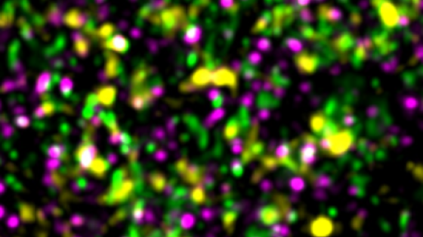
Banner image: Homer1 (green)- and synaptotagmin1/2 (magenta)-immunoreactive synapses express ‘eat-me’ signal phosphatidylserine as marked by PSVue643 (yellow) in the hippocampus of NL-F KI; Trem2 R47H KI mice. Credit: Javier Rueda-Carrasco, 10.15252/embj.2022113246
Video: Microglia eating synapses that are expressing the eat-me signal ePtdSer Credit: Sang-Eun Lee, 10.15252/embj.2022113246
Article published: 16 August 2023